Biomedical Optics
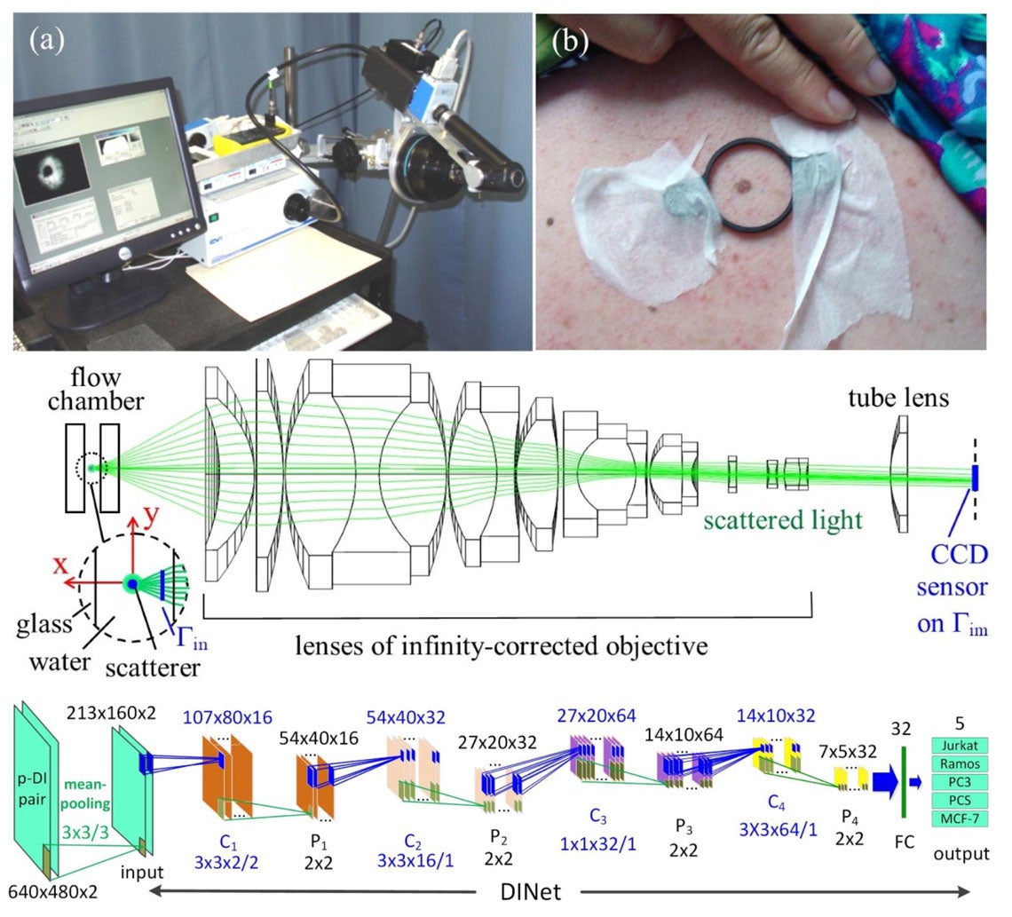
Xin-Hua Hu investigates the optical properties of biological samples and develops innovative instrumentation for the characterization of cells and turbid samples. Over the past decades he and Dr. Jun Q. Lu have developed in the Biomedical Laser Laboratory a laser-optics experimental research facility as well as a high-performance computing facility. Experimental measurements are carried out in the laser lab with in-house developed diffraction imaging flow cytometer, multispectral reflectance imaging systems, and multi-parameter spectrophotometry systems for image and spectroscopic data acquisition and analysis. Image processing and large scale numerical simulations have been actively pursued to clearly understand the correlations between cellular and turbid samples’ optical and morphological properties by imaging and measurement of scattered light signals. These research activities allow him to develop innovative approaches of acquiring big data on human cells and tissues with state-of-art machine learning algorithms and instrumentation for wide-ranged applications.

(a) A multispectral imaging system for multispectral imaging and diagnosis of cutaneous melanoma. (b) Preparation for image acquisition from an enrolled patient before surgical removal of a mole suspected of melanoma. (middle) The schematic of optical setup for diffraction imaging flow cytometry system with a microscope objective based imaging unit. (bottom) The dataflow graph of a deep learning neural network (DINet) developed for classification of 5 cell types based on cross-polarized diffraction image (p-DI) pair data.
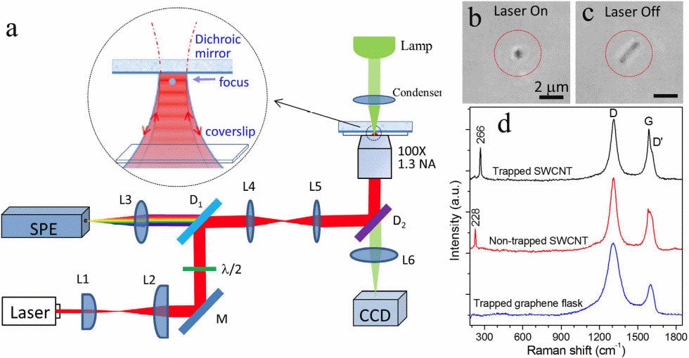
Yong-Qing Li studies the exciting field of biophotonics. Work in the Biomedical Optics Laboratory uses techniques such as optical tweezers and Raman spectroscopy and imaging, live-cell light microscopy, and atomic force microscopy to understand the fundamental biological processes of single cells and cellular heterogeneity. Projects include optical pulling of airborne particles and lifting of large objects by light, monitoring dynamic germination, outgrowth, and growth of single bacterial spores in nutrients and high pressure environment, and label-free surface enhanced Raman spectroscopy (SERS) for Ras proteins-based cancer diagnoses and therapy. There are both graduate- and undergraduate-level projects in the lab.

Raman tweezers is used for the stable optical trapping and characterization of nanoparticles and single cells. This figure’s source is part of a Nature collection on optical tweezers to celebrate the 2018 Nobel Prize in Physics.