Optically Stimulated Luminescence Laboratory
The Luminescence Laboratory is directed by Dr. Regina DeWitt, and is located in two separate, newly renovated laboratory areas in the Department of Physics. One area consists of a large room with two smaller side rooms (approx. 600 ft2) that is dedicated to instrument development. The second area (approx. 400 ft2) houses the luminescence dating facility. A dedicated beamline in the Accelerator Laboratory has been constructed to investigate the impact of heavy ion irradiation on the luminescence properties of minerals.
Luminescence Dating Facility
The laboratory is equipped with apparatuses for the detection/measurement of luminescence and its application to luminescence dating. This includes a Risø TL/OSL-DA-20 automated system with integrated 90Sr/Y beta and 241Am alpha radiation sources and all necessary computer control and analysis software. The instrument is equipped with built-in diodes for blue (470 nm), green (532nm), or IR-pulsed (875 nm) stimulation. Attachments for luminescence spectrometry as well as the detection of red emissions are currently under construction. The laboratory is further equipped with all necessary facilities for sample preparation, i.e., extractor hoods and dark rooms. Additionally, the laboratory has access to a high-purity Ge gamma spectrometer as well as a portable LaBr gamma spectrometer.
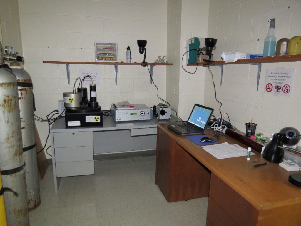
Risø TL/OSL-DA-20 automated system.
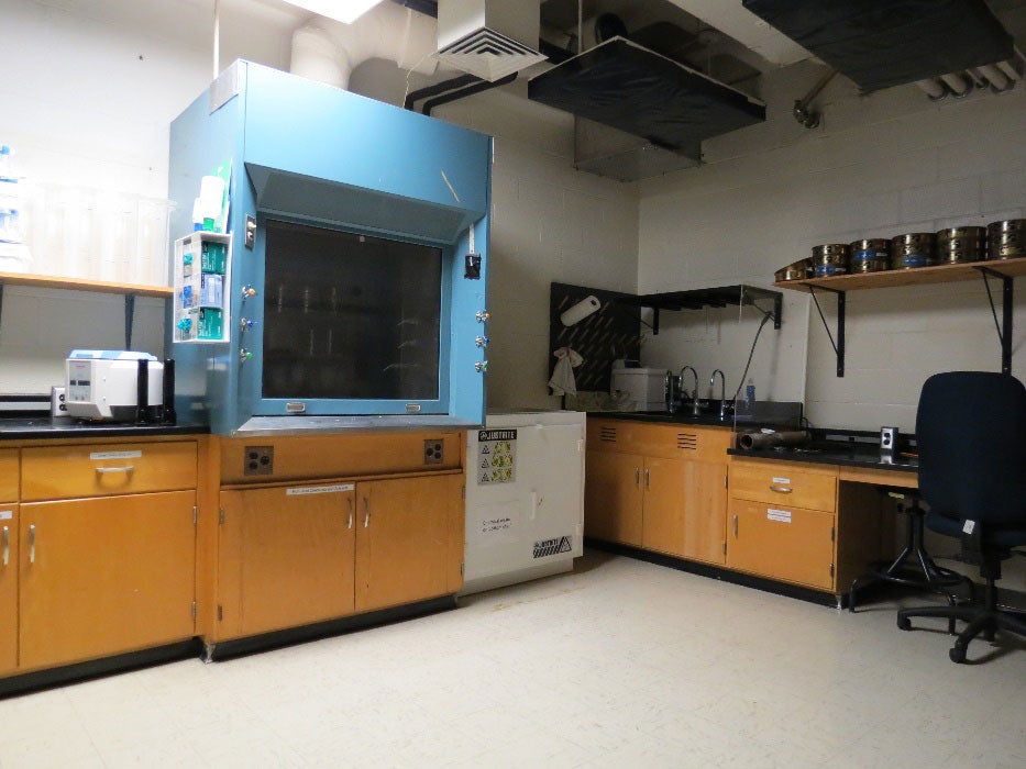
Right side of the sample preparation room with fume hood and sink.
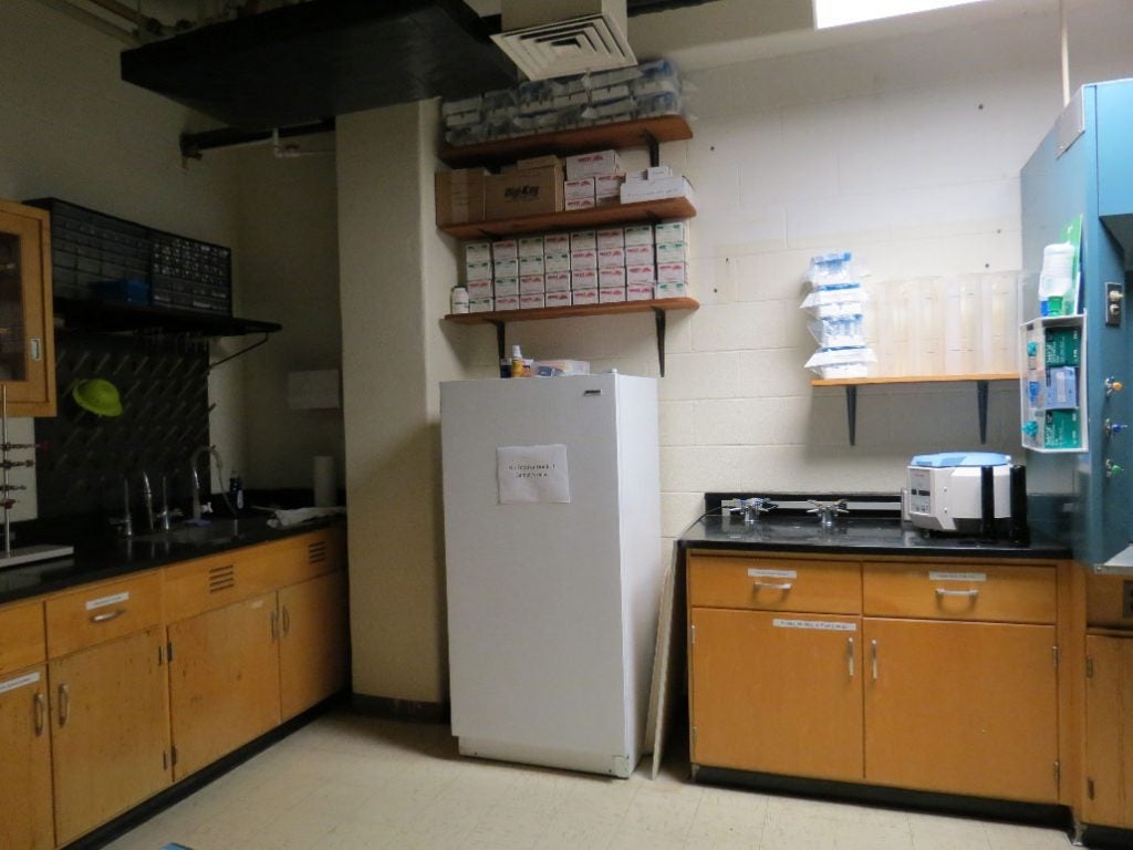
Left side of the sample preparation room with second sink and freezer for cold storage.
Instrument Development: LuCIDD
The Luminescence instrument with Confocal and Imaging units for Dating and Dosimetry (LuCIDD) is being developed and tested as a novel device for luminescence imaging with funding from the NSF MRI Program. The instrument consists of two units: One is based on a confocal microscope, where a narrow laser spot is scanned over the surface and the signal is detected with a photomultiplier tube. The confocal setup blocks signals emitted by deeper, unbleached grains. The unit is equipped with blue, green, red, and IR lasers for stimulation. The system is configured to measure the UV signal, but can also be modified to measure other emissions such as blue. A typical laser stimulation spot size is approximately 50-100 μm. The second unit consists of a CCD-based system with a ring of blue and IR LEDs for stimulation. Both units allow temperature control of the sample (up to 400 °C). An X-ray source enables continuous or pulsed mode irradiation.

Confocal unit of LuCIDD.
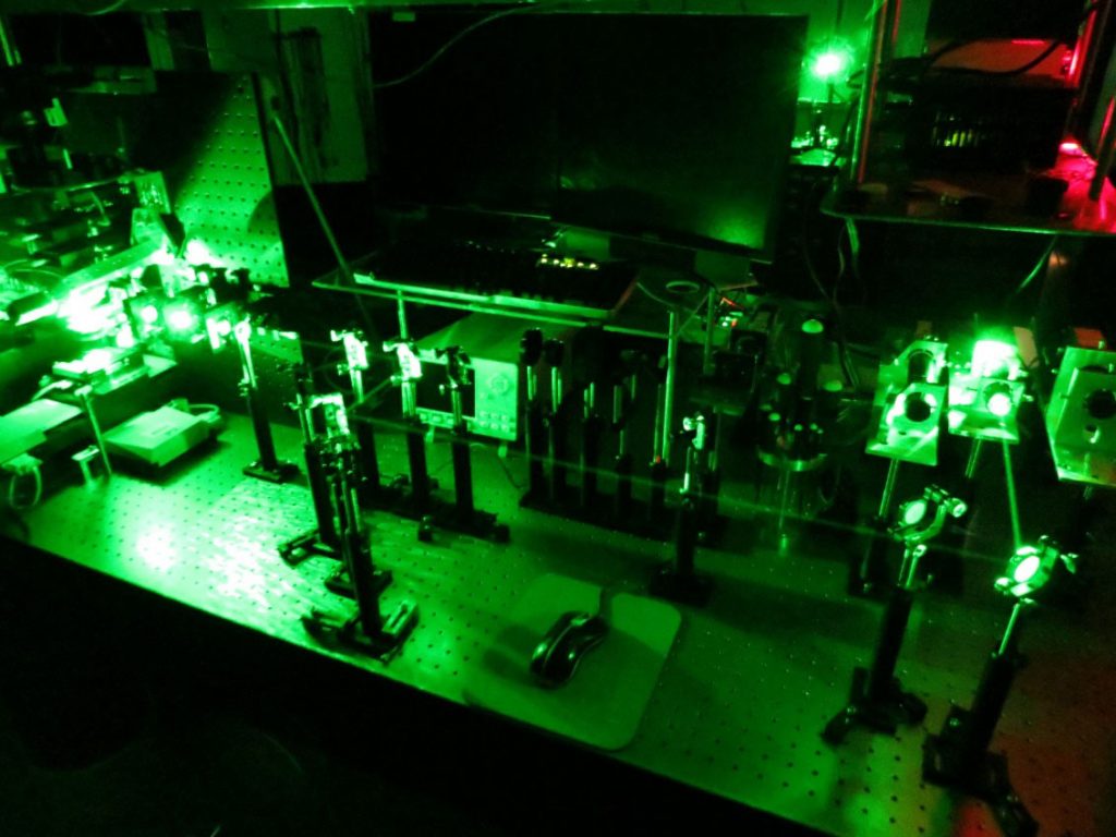
Optical path of LuCIDD.
Instrument Development: ODIN
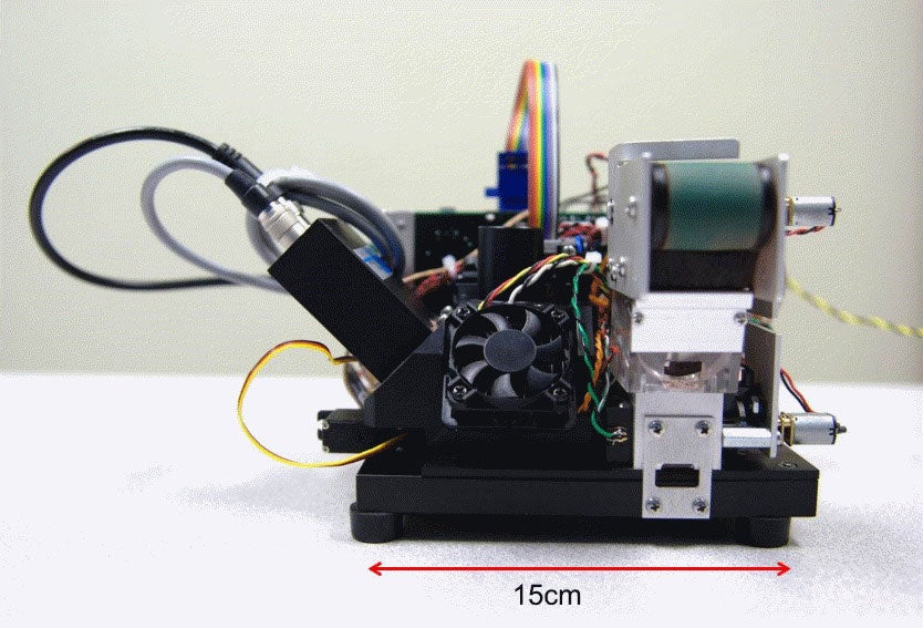
Optical Dating Instrument (ODIN).
Accelerator Beamline
One of our accelerator beamlines is dedicated to luminescence studies. The beamline consists of a beampipe with vacuum pumps, gauges, beam profile monitor, and Faraday cup. The sample chamber and transfer port can be sealed off via a gate valve. A black curtain enclosure ensures that samples can be removed from the chamber after irradiation without light exposure. Up to six samples can be mounted on a sample ladder for irradiation.
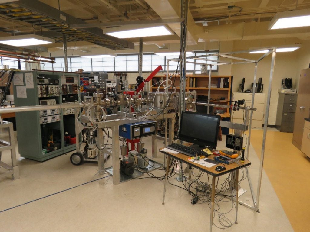
Luminescence beamline prior to attachment of the blackout curtain.
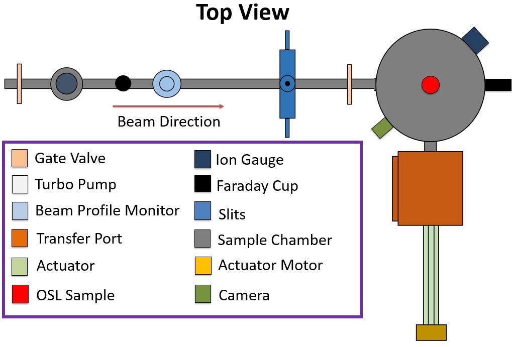
Schematic of the beamline, sample chamber, and actuator with key indicating specific components.